
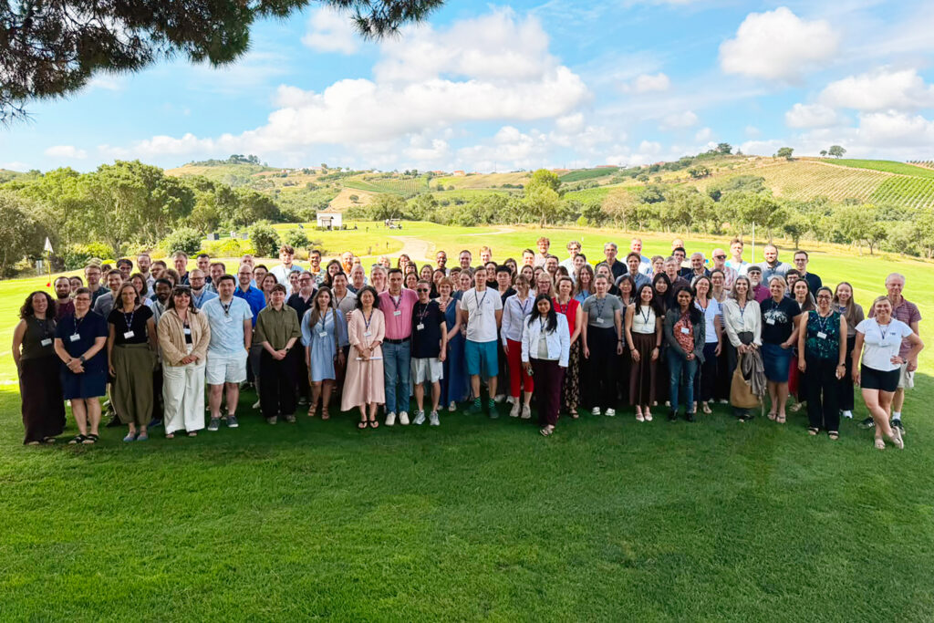
International Conference on the Cell Biology of Single-Celled Organisms
Professor Marc Bramkamp from Kiel University co-organises conference in Lisbon on new cell biology methods, supported by the Kiel Life Science research priority area.
From 13 to 16 July, the Prokaryotic Cell Biology conference (Prokaryotes-including bacteria and archaea) takes place in the Portuguese capital of Lisbon. The conference is co-organised by Professor Marc Bramkamp from the Institute of General Microbiology at Kiel University. More than 100 participants gather at this major international meeting of microbiologists to exchange ideas on current developments in cell biology, advances in imaging technologies, and other innovative analytical methods. The conference is supported by the Federation of European Microbiological Societies (FEMS) and Kiel University’s research priority area Kiel Life Science (KLS). It is chaired by Professor Jie Xiao of Johns Hopkins University, Professor Heath Murray of Newcastle University, Professor Ethan Garner of Harvard University, and Professor Marc Bramkamp, member of the CRC 1182 in Kiel.
Advances in methodology and imaging open up new perspectives in cell biology
Over the past two decades, research in the life sciences has increasingly focused on the fundamental functioning of single-celled organisms such as bacteria and archaea. This development has been driven primarily by advances in microscopy, genome sequencing, and bioinformatics. New approaches that combine molecular information with its spatial distribution within cells and organisms are expanding the possibilities for studying biological processes in their native context. This research approach, also known as spatial biology, enables, for the first time, a comprehensive mechanistic understanding of how a living cell functions.
“Major technological innovations have significantly expanded the range of organisms that can be studied, enabling us to analyse and compare processes such as growth, morphogenesis, and cell division across numerous single-celled organisms. This allows us to gain new insights into the molecular mechanisms responsible for cellular function in detail. In turn, this helps us to better understand the fundamental biological organisation of life as a whole,” emphasises Bramkamp, who heads the Microbial Biochemistry and Cell Biology research group and the Kiel University Light and Electron Microscopy Bioscience Facility (KLEM) at Kiel University.
As part of the conference programme, participants will explore six key topics through around 40 lectures, poster sessions, and dedicated events for early-career researchers: Advances in Nanoimaging, Cell Envelope Dynamics and Division, Cell Growth and the Cell Cycle, Cell Shape and Morphogenesis, Genome Organisation, Replication and Expression, and Spatial Organisation of Cellular Structures. Highlights include plenary lectures by Professor Christine Jacobs-Wagner (Stanford University), Professor Jeff Errington (University of Sydney), and Professor Jan Löwe (University of Cambridge), as well as presentations by many outstanding early-career scientists, including Dr Benjamin Springstein (Vienna) and Anne-Marie Wehenkel (Paris).
“A detailed understanding of the molecular mechanisms underlying cell organisation and cell division is a prerequisite for being able to intervene in these processes in a targeted way in the future. The knowledge gained will therefore open up new long-term perspectives—for example, for combating microbial pathogens or for the development and control of microbial systems in biotechnology,” says Bramkamp.
Strengthening innovative life science methods at Kiel University
Kiel University’s contributions to the current conference in Lisbon also reflect the university’s successful efforts to establish and consolidate new biological imaging methods. A large proportion of the research presented at the conference is based on innovative analytical techniques that have also been established at Kiel University. Among these is the highly specialised Kiel University Light and Electron Microscopy Bioscience Facility (KLEM), which operates within the Central Microscopy Unit (ZM) of the Centre for Biology. The facility houses, among other instruments, confocal laser scanning microscopes, a super-resolution microscope for applications such as single-molecule localisation microscopy, transmission electron microscopes (TEM), and a scanning electron microscope (SEM). Together with state-of-the-art bioinformatics facilities and an advanced genome sequencing centre, these resources provide Kiel University researchers with cutting-edge methods for life science research.
“We have succeeded in establishing a broad portfolio of innovative, state-of-the-art microscopy technologies over the past few years that can easily compete at the international level. This gives us in Kiel an important key to addressing fundamental questions in the life sciences at the highest level. We also want to make these capabilities available to other users through Kiel University’s research priority area Kiel Life Science and beyond,” Bramkamp concludes.
More information:
Prokaryotic Cell Biology-Conference, Lisbon, 13 -16 July:
fusion-conferences.com/conference/198
Microbial Biochemistry and Cell Biology (Bramkamp Lab)
Institute for General Microbiology, Kiel University:
www.bramkamplab.org
Kiel University Light and Electron Microscopy Bioscience Facility (KLEM)
Department of Biology, Kiel University:
www.biologie.uni-kiel.de/zm/de/dfg-risources-klem